Our instability series includes the following views:
- True AP
- Axillary Lateral View
- Stryker Notch View
- Westpoint View
Each of these is listed below with examples and a description of how to shoot it.
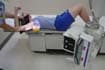
True AP
Position: Patient erect with affected side against Bucky, turn patient 30-35 degrees
Tube: Perpendicular
Demonstrates: Glenohumeral joint space
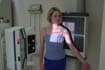
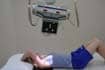
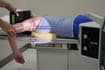
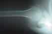
Axillary Lateral
Position: Patient supine with small sponge under shoulder, humerus abducted to 90 degrees, elbow bent 90 degrees and perpendicular to table
Tube: Central ray bisecting angle of humerus and body
Demonstrates:
Glenohumeral joint
Coracoid process pointing anteriorly
Lessor tuberosity in profile
AC joint, acromion, and end of clavicle project through
humeral head

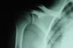
Stryker Notch View
Position: Erect or supine, with hand on back of head
Tube: 10 degrees cephalic to the coracoid
Demonstrates:
Hill-Sachs defects
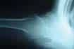
Westpoint View
Position: Prone with sponge supporting affected shoulder, abduct arm 90 degrees, bend elbow and hang forearm off the table
Tube: Central ray is double angled – 25 degrees from horizontal and 25 degrees medially
Demonstrates:
Bony abnormalities of the glenoid rim
Instability